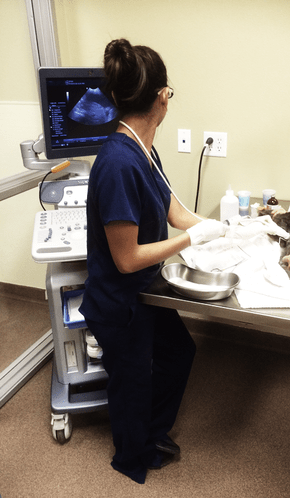
ULTRASOUND

Although humans and animals are different in many ways, some advances in human medicine are also very useful for veterinary patients. One of these advances, diagnostic ultrasound, has proven to be a powerful tool in veterinary medicine. As a practice, one of our goals is to offer state-of-the-art medicine and diagnostic testing; so we are pleased to offer ultrasound services as a means of providing a higher level of quality care to our patients.
Ultrasonography is a type of diagnostic technique that uses ultrasound waves to produce an imaging study. This means that when we perform ultrasonography, we can see internal images of the patient’s body. Unlike some other imaging studies, like x-rays, ultrasonography does not use radiation. Instead, ultrasonography uses high-frequency sound (ultrasound) waves to create a picture of what is inside your pet’s body. Ultrasonography is a completely non-invasive, painless way to diagnose and evaluate many common diseases.
An ultrasound machine generates ultrasound waves. The machine is connected to a small probe that is held gently against your pet’s skin. The probe sends out painless ultrasound waves that bounce off of structures (for example, organs) in your pet’s body and return to a sensor inside the ultrasound machine. The ultrasound equipment collects these reflected “echoes” and uses them to generate images that are viewable on a screen. Ultrasound waves can generate excellent images of abdominal organs, including the liver, spleen, gallbladder, and kidneys. It is also useful for assessing fetal health and monitoring pregnancy in breeding animals, and it can help us diagnose and stage (determine the severity of) some forms of cancer.
Because ultrasound images are produced in real time, this technology can be used to evaluate the heart as it beats. This can help us detect abnormalities in the motion of heart valves, blood flow through the heart, and contractions of the heart muscle. It can also be used to assess the heart for defects. As we strive to provide our patients with the highest quality medicine and diagnostic testing, we are pleased to offer ultrasound as one of our diagnostic capabilities.

Lindl Bylicki, DVM, DACVR
Ultrasound/Radiology Specialist
Britany Lindl Bylicki is originally from Livermore, CA where most of her family still resides. She received a BS from UC Santa Barbara and graduated with high honors in Biopsychology in 2005. She returned to Northern California to attend The School of Veterinary Medicine at UC Davis and graduated in 2010. After that, it was back to Southern California for a rotating small animal internship at the Veterinary Medical and Surgical Group in Ventura, CA. After a year at VMSG, she returned to UC Davis for a four-year residency in diagnostic imaging. Britany became a Diplomate of the American College of Veterinary Radiologist in 2014.
She is an author of several publications in the areas of carotid body tumors, the canine dorsal tracheal membrane, chondro-osseous respiratory epithelial adenomatoid hamartomas, and CT pneumocolonography. She serves as a peer reviewer for the Journal of the American Veterinary Medical Association and the Journal of Veterinary Research.
She currently lives in West Sacramento with her husband and their Border Collie mix. They enjoy hiking and backpacking, snowboarding, and finding new beer to try. Britany is an active member of the River City Rowing Club’s Open Women’s team and travels the country as a competitive rower.
